|
Sino Biological
rabbit anti bira  Rabbit Anti Bira, supplied by Sino Biological, used in various techniques. Bioz Stars score: 93/100, based on 1 PubMed citations. ZERO BIAS - scores, article reviews, protocol conditions and more https://www.bioz.com/result/rabbit anti bira/product/Sino Biological Average 93 stars, based on 1 article reviews
rabbit anti bira - by Bioz Stars,
2026-06
93/100 stars
|
Buy from Supplier |
|
Proteintech
rabbit polyclonal anti luzp1 antibody  Rabbit Polyclonal Anti Luzp1 Antibody, supplied by Proteintech, used in various techniques. Bioz Stars score: 93/100, based on 1 PubMed citations. ZERO BIAS - scores, article reviews, protocol conditions and more https://www.bioz.com/result/rabbit polyclonal anti luzp1 antibody/product/Proteintech Average 93 stars, based on 1 article reviews
rabbit polyclonal anti luzp1 antibody - by Bioz Stars,
2026-06
93/100 stars
|
Buy from Supplier |
|
Bethyl
luzp1 antibody  Luzp1 Antibody, supplied by Bethyl, used in various techniques. Bioz Stars score: 92/100, based on 1 PubMed citations. ZERO BIAS - scores, article reviews, protocol conditions and more https://www.bioz.com/result/luzp1 antibody/product/Bethyl Average 92 stars, based on 1 article reviews
luzp1 antibody - by Bioz Stars,
2026-06
92/100 stars
|
Buy from Supplier |
|
Proteintech
mouse anti gapdh Mouse Anti Gapdh, supplied by Proteintech, used in various techniques. Bioz Stars score: 96/100, based on 1 PubMed citations. ZERO BIAS - scores, article reviews, protocol conditions and more https://www.bioz.com/result/mouse anti gapdh/product/Proteintech Average 96 stars, based on 1 article reviews
mouse anti gapdh - by Bioz Stars,
2026-06
96/100 stars
|
Buy from Supplier |
|
Bethyl
rabbit anti ccp110 Rabbit Anti Ccp110, supplied by Bethyl, used in various techniques. Bioz Stars score: 93/100, based on 1 PubMed citations. ZERO BIAS - scores, article reviews, protocol conditions and more https://www.bioz.com/result/rabbit anti ccp110/product/Bethyl Average 93 stars, based on 1 article reviews
rabbit anti ccp110 - by Bioz Stars,
2026-06
93/100 stars
|
Buy from Supplier |
|
Proteintech
anti odf2 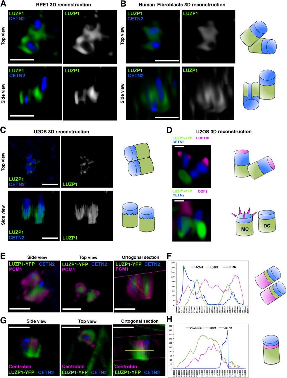 Anti Odf2, supplied by Proteintech, used in various techniques. Bioz Stars score: 93/100, based on 1 PubMed citations. ZERO BIAS - scores, article reviews, protocol conditions and more https://www.bioz.com/result/anti odf2/product/Proteintech Average 93 stars, based on 1 article reviews
anti odf2 - by Bioz Stars,
2026-06
93/100 stars
|
Buy from Supplier |
|
Proteintech
rest Rest, supplied by Proteintech, used in various techniques. Bioz Stars score: 94/100, based on 1 PubMed citations. ZERO BIAS - scores, article reviews, protocol conditions and more https://www.bioz.com/result/rest/product/Proteintech Average 94 stars, based on 1 article reviews
rest - by Bioz Stars,
2026-06
94/100 stars
|
Buy from Supplier |
|
Proteintech
anti cep97 Anti Cep97, supplied by Proteintech, used in various techniques. Bioz Stars score: 93/100, based on 1 PubMed citations. ZERO BIAS - scores, article reviews, protocol conditions and more https://www.bioz.com/result/anti cep97/product/Proteintech Average 93 stars, based on 1 article reviews
anti cep97 - by Bioz Stars,
2026-06
93/100 stars
|
Buy from Supplier |
|
Cell Signaling Technology Inc
myc Myc, supplied by Cell Signaling Technology Inc, used in various techniques. Bioz Stars score: 95/100, based on 1 PubMed citations. ZERO BIAS - scores, article reviews, protocol conditions and more https://www.bioz.com/result/myc/product/Cell Signaling Technology Inc Average 95 stars, based on 1 article reviews
myc - by Bioz Stars,
2026-06
95/100 stars
|
Buy from Supplier |
|
Cell Signaling Technology Inc
rabbit anti pcm1 Rabbit Anti Pcm1, supplied by Cell Signaling Technology Inc, used in various techniques. Bioz Stars score: 86/100, based on 1 PubMed citations. ZERO BIAS - scores, article reviews, protocol conditions and more https://www.bioz.com/result/rabbit anti pcm1/product/Cell Signaling Technology Inc Average 86 stars, based on 1 article reviews
rabbit anti pcm1 - by Bioz Stars,
2026-06
86/100 stars
|
Buy from Supplier |
|
GeneTex
mouse anti-cep164  Mouse Anti Cep164, supplied by GeneTex, used in various techniques. Bioz Stars score: 90/100, based on 1 PubMed citations. ZERO BIAS - scores, article reviews, protocol conditions and more https://www.bioz.com/result/mouse anti-cep164/product/GeneTex Average 90 stars, based on 1 article reviews
mouse anti-cep164 - by Bioz Stars,
2026-06
90/100 stars
|
Buy from Supplier |
Image Search Results
Journal: eLife
Article Title: LUZP1, a novel regulator of primary cilia and the actin cytoskeleton, is a contributing factor in Townes-Brocks Syndrome
doi: 10.7554/eLife.55957
Figure Lengend Snippet: ( A ) Western blot of inputs or GFP-Trap pulldowns performed in HEK 293FT cells transfected with SALL1 275 -YFP (lanes 1 and 6), SALL1 FL -YFP (lanes 2 and 7), YFP alone (lanes 3 and 8), SALL1 275 -YFP together with SALL1 FL -2xHA (SALL1 FL -HA; lanes 4 and 9) or SALL1 FL -HA together with YFP alone (lanes 5 and 10). Specific antibodies (LUZP1, actin, SALL1) were used as indicated. Numbers under LUZP1 panel result from dividing band intensities of each pulldown by their respective input levels. One asterisk indicates BirA*-SALL1 FL or SALL1 FL -YFP, one black arrowhead SALL1 275 -YFP and two black arrowheads YFP alone. Molecular weight markers (kDa) are shown to the right. Actin was used as loading control. Blots shown are representative of three independent experiments. ( B ) Western blot analysis of RPE1 cells transfected with TbID-LUZP1 or TbID alone. Specific antibodies (BirA, LUZP1 and GAPDH) were used as indicated. Anti-BirA antibody detected the fusion form of TbID-LUZP1 (two asterisks) or TbID (three asterisks). LUZP1 antibodies detected endogenous LUZP1 (two empty arrowheads) and TbID-LUZP1 (two arrowheads). Molecular weight markers are shown to the right. ( C ) In RPE1 cells transfected with TbID-LUZP1, streptavidin (green) localizes to the centrosome labelled with Pericentrin (PCNT, magenta) or actin fibers labelled with phalloidin (Phall, magenta). No biotinylation in microtubules, labelled by βTubulin (βTub, magenta), was observed. By contrast, cells transfected with TbID show streptavidin labeling in nucleus and cytoplasm. Green channels are shown in black and white. Arrowheads indicate the centrioles. Note that basal actin stress fibers are less evident when apical centrioles are in focus.
Article Snippet: Antibodies used: rabbit anti-LUZP1 (Proteintech, 1:1,000) for and rabbit anti-LUZP1 (Sigma HPA028506, 1:1,000) for the rest of the experiments, rabbit anti-CCP110 (Proteintech, 1:1,000), rabbit anti-CEP97 (Proteintech, 1:1,000), mouse anti-GFP (Roche, 1:1,000), mouse anti-GAPDH (Proteintech, 1:1,000), mouse anti-FLNA (Merck, 1:1,000),
Techniques: Western Blot, Transfection, Molecular Weight, Labeling
Journal: eLife
Article Title: LUZP1, a novel regulator of primary cilia and the actin cytoskeleton, is a contributing factor in Townes-Brocks Syndrome
doi: 10.7554/eLife.55957
Figure Lengend Snippet: ( A ) Representative western blot of Control and TBS 275 total cell lysates treated or not with MG132. A specific antibody detected endogenous LUZP1, and GAPDH was used as loading control. ( B ) Graphical representation of the fold changes of LUZP1/GAPDH ratios obtained in ( A ) for of Control (blue dots) and TBS 275 (orange dots) treated (+) or not (-) with the proteasome inhibitor MG132. Note the increase of LUZP1 until reaching control levels in TBS 275 cells upon MG132 treatment. ( C ) Representative western blot of 293 WT and 293 335 total cell lysates treated or not with MG132. A specific antibody against LUZP1 detected endogenous LUZP1, and GAPDH was used as loading control. ( D ) Graphical representation of the fold changes of LUZP1/GAPDH ratios obtained in panel C for 293 WT (blue dots) and 293 335 (orange dots) treated (+) or not (-) with MG132. Note that LUZP1 in 293 335 reaches control levels with MG132 treatment. ( E ) Representative western blot of total lysates of HEK 293FT cells transfected with SALL1 275 -YFP (lanes 1 and 3) or YFP alone (lanes 2 and 4) treated (+) or not (-) with MG132. Specific antibodies against LUZP1, GFP and GAPDH were used. One black arrowhead indicates SALL1 275 -YFP, two back arrowheads YFP alone. ( F ) Graphical representation of the fold changes of LUZP1/GAPDH ratios obtained in ( E ) for HEK 293FT cells transfected with SALL1 275 -YFP (orange dots) or YFP alone (blue dots) treated (+) or not (-) with MG132. Note that LUZP1 increases in the presence of MG132 when SALL1 275 -YFP was transfected. Data from at least three independent experiments pooled together are shown. P -values were calculated using two-tailed unpaired Student´s t-test. ( G ) Immunofluorescence micrographs of RPE1 cells treated (+MG132) or not (-MG132) with proteasome inhibitor showing LUZP1 associated with the cytoskeleton (upper panels) or in the centrosome (lower panels). Antibodies against endogenous LUZP1 (green), Centrin-2 (CETN2, blue) and CEP164 (magenta) were used. DAPI labelled the nuclei (blue). Single green channels are shown in black and white. Note the overall increase of LUZP1 upon MG132 treatment. Scale bar 10 µm (cytoskeleton panels) or 0.5 µm (centrosome panels). Imaging was performed using widefield fluorescence microscopy (Zeiss Axioimager D1, 63x objective). ( H ) Western blot analysis of input and biotin pulldown (PD) of HEK 293FT cells transfected with CMV-LUZP1-YFP and BioUb or BirA alone treated (+) or not (-) with MG132. Specific antibodies (GFP, GAPDH, Biotin) were used as indicated. Numbers under GFP panel are the result of dividing each biotin PD band intensity by the respective input band intensity and normalize them to lane 1. Molecular weight markers in kDa are shown to the right. Two asterisks indicate monoubiquitinated LUZP1.
Article Snippet: Antibodies used: rabbit anti-LUZP1 (Proteintech, 1:1,000) for and rabbit anti-LUZP1 (Sigma HPA028506, 1:1,000) for the rest of the experiments, rabbit anti-CCP110 (Proteintech, 1:1,000), rabbit anti-CEP97 (Proteintech, 1:1,000), mouse anti-GFP (Roche, 1:1,000), mouse anti-GAPDH (Proteintech, 1:1,000), mouse anti-FLNA (Merck, 1:1,000),
Techniques: Western Blot, Transfection, Two Tailed Test, Immunofluorescence, Imaging, Fluorescence, Microscopy, Molecular Weight
Journal: The Journal of Biological Chemistry
Article Title: Activating transcription factor 4 (ATF4) promotes skeletal muscle atrophy by forming a heterodimer with the transcriptional regulator C/EBPβ
doi: 10.1074/jbc.RA119.012095
Figure Lengend Snippet: Identification of proteins that interact with the ATF4 bZIP domain in mouse skeletal muscle fibers. A, control and ATF4 bZIP pulldown samples from two independent, time-separated experiments were obtained as described in D and then subjected to MS and a data analysis workflow that is fully detailed under “Experimental procedures” and the supporting information . The table in A summarizes the mass spectrometric data for ATF4 and the high-confidence ATF4-interacting proteins that were identified in the ATF4 bZIP pulldown samples. For proteins with only one peptide identified, the corresponding MS/MS were manually inspected (also see under “Experimental procedures” and Fig. S2 ). B, tandem mass spectrum (MS/MS) from a representative C/EBPβ peptide VLELTAENER (SwissProt accession P28033) spanning residues 254–263 with the precursor ion at m/z 587.31 2 + identified in the ATF4 bZIP affinity-enrichment sample. C, left , quantification of C/EBPβ peptide VLELTAENER using DIA MS2-based quantification; panels show extracted ion chromatograms for the MS2 fragment ions in the control and ATF4 bZIP affinity enrichment sample. Right , MS/MS spectral library “simulation” indicating relative fragment ion distribution and calculated peak areas under the curve for the C/EBPβ peptide VLELTAENER in control and ATF4 bZIP affinity enrichment samples. D, mapping of VLELTAENER to the crystal structure of the ATF4 and C/EBPβ bZIP domains . E, TA muscle fibers of 12 mice were transfected with 20 μg of empty TAP plasmid (one TA per mouse) or 20 μg of ATF4 bZIP TAP plasmid (the contralateral TA in each mouse). Seven days post-transfection, bilateral TA muscles were harvested and used to prepare pooled protein extracts from each of the two groups of skeletal muscles (control and ATF4 bZIP). The pooled protein extracts were then subjected to pulldown with anti-FLAG magnetic beads, followed by SDS-PAGE and immunoblot analysis using HRP-conjugated mouse monoclonal anti-FLAG IgG ( upper panel ), mouse monoclonal anti-C/EBPβ IgG, rabbit polyclonal anti-C/EBPδ antibody, rabbit monoclonal anti-C/EBPα IgG, rabbit polyclonal anti-MAF antibody, and rabbit polyclonal anti-LUZP1 antibody, as indicated. Asterisk in the anti-C/EBPβ immunoblot denotes a nonspecific cross-reacting protein.
Article Snippet:
Techniques: Control, Tandem Mass Spectroscopy, Transfection, Plasmid Preparation, Muscles, Magnetic Beads, SDS Page, Western Blot

Journal: The Journal of Biological Chemistry
Article Title: Activating transcription factor 4 (ATF4) promotes skeletal muscle atrophy by forming a heterodimer with the transcriptional regulator C/EBPβ
doi: 10.1074/jbc.RA119.012095
Figure Lengend Snippet: Development of RNAi constructs that target ATF4 interactors in mouse skeletal muscle fibers. Mouse TA muscles were co-transfected with 10 μg of cDNA (C/EBPα plasmid in A ; C/EBPβ plasmid in B ; C/EBPγ plasmid in C ; C/EBPδ plasmid in D ; MAF plasmid in E ; LUZP1–FLAG plasmid in F ; and BRD4–FLAG plasmid in G ) plus either 20 μg of nontargeting control RNAi plasmid or 20 μg of RNAi plasmid targeting C/EBPα ( A ), C/EBPβ ( B ), C/EBPγ ( C ), C/EBPδ ( D ), MAF ( E ), LUZP1 ( F ), or BRD4 ( G ), as indicated. Seven days post-transfection, bilateral TA muscles were harvested for SDS-PAGE and immunoblot analysis using rabbit monoclonal anti-C/EBPα IgG ( A ), mouse monoclonal anti-C/EBPβ IgG ( B ), rabbit polyclonal anti-C/EBPγ antibody ( C ), rabbit polyclonal anti-C/EBPδ antibody ( D ), rabbit polyclonal anti-MAF antibody ( E ), and HRP-conjugated mouse monoclonal anti-FLAG IgG ( F and G ). Membranes were stained with Ponceau S to confirm equal loading.
Article Snippet:
Techniques: Construct, Muscles, Transfection, Plasmid Preparation, Control, SDS Page, Western Blot, Staining

Journal: The Journal of Biological Chemistry
Article Title: Activating transcription factor 4 (ATF4) promotes skeletal muscle atrophy by forming a heterodimer with the transcriptional regulator C/EBPβ
doi: 10.1074/jbc.RA119.012095
Figure Lengend Snippet: In vivo RNAi-based phenotypic screen of ATF4 interactors in mouse skeletal muscle fibers. A, one TA muscle per mouse was transfected with 5 μg of empty pcDNA plasmid plus 2 μg of eGFP plasmid, and the contralateral TA in each mouse was transfected with 5 μg of ATF4–FLAG plasmid plus 2 μg of eGFP plasmid. B–H , one TA per mouse was transfected with 5 μg of ATF4–FLAG plasmid plus 10 μg of control RNAi plasmid, and the contralateral TA in each mouse was transfected with 5 μg of ATF4–FLAG plasmid plus 10 μg of RNAi plasmid targeting C/EBPα ( B ), C/EBPβ ( C ), C/EBPγ ( D ), C/EBPδ ( E ), MAF ( F ), LUZP1 ( G ), or BRD4 ( H ), as indicated. A–H , 7 days post-transfection, bilateral TAs were harvested for histological analysis. Left panels : size distribution of >4700 transfected muscle fibers from ≥5 muscles per condition. Right panels : each data point represents the mean fiber diameter from one TA (>550 fibers measured per muscle), and horizontal bars denote average of the means. p values were determined with paired t tests.
Article Snippet:
Techniques: In Vivo, Transfection, Plasmid Preparation, Control, Muscles
Journal: PNAS Nexus
Article Title: Death-associated protein kinase 3 modulates migration and invasion of triple-negative breast cancer cells
doi: 10.1093/pnasnexus/pgae401
Figure Lengend Snippet: IP–MS identifies that LUZP1 is one of the top interacting proteins of DAPK3. A) Schematic representation of IP–MS workflow using a DAPK3 antibody in SUM159 control and DAPK3 KO #3 cells. B) Top 10 MF terms of GO overrepresentation analysis with a total of 73 DAPK3-interacting proteins identified from IP–MS analysis. P -values are determined by the overrepresentation analysis. C) Top 5 interacting proteins of DAPK3 identified from IP–MS analysis. The proteins are ranked based on the ratio of iBAQ levels in DAPK3 KO #3 cells compared with control cells. The full list of proteins in the DAPK3 immunoprecipitants is shown in Table . D) Immunoblotting shows the protein levels of LUZP1 and DAPK3 in whole-cell lysates and co-IP samples of anti-DAPK3 antibody obtained from SUM159 control and DAPK3 KO #3 cells. The representative image is based on two independent experiments. E) Immunoblotting shows the protein levels of LUZP1 and DAPK3 in whole-cell lysates and co-IP samples of antiLUZP1 antibody obtained from SUM159 control and LUZP1 KO #6 cells. The representative image is based on two independent experiments. F) Immunoblotting shows the protein levels of LUZP1 and HA-tagged DAPK3 in whole-cell lysates and co-IP samples of anti-HA antibody obtained from SUM159 DAPK3 KO #3 cell, DAPK3 KO #3 cells with HA-tagged DAPK3 WT overexpression, and DAPK3 KO #3 cells with HA-tagged DAPK3 ΔLZ overexpression. The representative image is based on two independent experiments.
Article Snippet: For IP–immunoblotting, 1 mg of lysates was incubated with 1 μg DAPK3 antibody (Santa Cruz Biotechnology, sc-514223), or 1 μg
Techniques: Protein-Protein interactions, Control, Western Blot, Co-Immunoprecipitation Assay, Over Expression

Journal: PNAS Nexus
Article Title: Death-associated protein kinase 3 modulates migration and invasion of triple-negative breast cancer cells
doi: 10.1093/pnasnexus/pgae401
Figure Lengend Snippet: LUZP1 knockout markedly decreased DAPK3 protein levels, but did not change DAPK3 mRNA levels. A) Immunoblotting shows the protein levels of LUZP1 and DAPK3 in SUM159 control, DAPK3 KO #3, DAPK3 KO #6, LUZP1 KO #6, and LUZP1 KO #8 cells. GAPDH serves as a loading control. The representative image is based on three independent experiments. B), C) mRNA expression levels of DAPK3 (B) and LUZP1 (C) in SUM159 control, DAPK3 KO #3, DAPK3 KO #6, LUZP1 KO #6, and LUZP1 KO #8 cells. Values were normalized to GAPDH mRNA, and relative expression level was calculated as fold change to SUM159 control cells. Data represent the mean ± SEM from three independent experiments. NS, not significant, **** P < 0.0001, *** P < 0.001, * P < 0.05, two-way ANOVA with Dunnett's multiple comparisons test. D) Immunoblotting shows the protein levels of LUZP1 and DAPK3 in SUM159 control, LUZP1 KO #6, and LUZP1 KO #8 cells with or without MG-132 treatment (10 μM for 16 h). GAPDH serves as a loading control. The representative image is based on three independent experiments. E) Schematic presentation of the functional role of DAPK3 in TNBC cells, which involves interacting with LUZP1, to drive a mesenchymal cell state.
Article Snippet: For IP–immunoblotting, 1 mg of lysates was incubated with 1 μg DAPK3 antibody (Santa Cruz Biotechnology, sc-514223), or 1 μg
Techniques: Knock-Out, Western Blot, Control, Expressing, Functional Assay
Journal: bioRxiv
Article Title: LUZP1, a novel regulator of primary cilia and the actin cytoskeleton, is altered in Townes-Brocks Syndrome
doi: 10.1101/721316
Figure Lengend Snippet: ( A, B, C ) 2D images of a 3D reconstruction of immunofluorescence micrographs of LUZP1 (green) and Centrin-2 (CETN2, blue) in RPE1 cells ( A ), control fibroblasts (ESCTRL#2) ( B ) or U2OS cells ( C ). ( D ) 2D sections of U2OS cells overexpressing GFP-LUZP1 (green) stained with antibodies against CCP110 (upper panel, magenta) or ODF2 (lower panel, magenta) and CETN2 (blue). ( E, G ) 3D immunofluorescence micrographs of U2OS cells overexpressing LUZP1-YFP (green) stained with antibodies against PCM1 ( E ) or centrobin ( G ) in magenta and CETN2 (blue). Purple lines indicate the orthogonal cuts of the confocal z-stacks sections; yellow lines indicate the quantification point in ( F ) and ( H ). ( F, H ) Plot profile of the orthogonal section in ( E ) or ( G ) showing LUZP1-YFP (green), PCM1 or centrobin (magenta) and CETN2 (blue) intensities along the yellow lines in E and G , from left to right. Schematic representation of LUZP1 localization at the centrosome was modelled according to their respective micrographs in ( A-G ). Scale bar, 1 µm ( A-D ) or 0.5 µm ( D, E, G ). Imaging was performed using confocal microscopy (Leica SP8, 63x objective). Lightning software (Leica) was applied. The following figure supplement is available for : . Centrosomal localization of LUZP1 along the cell cycle. Figure 2-Supplementary video 1. LUZP1 localization in the centrosome.
Article Snippet: Other antibodies include, anti Centrin-2 (CETN2, Biolegend, 1:160), anti-LUZP1 (Sigma, 1:100), anti-LUZP1 (Proteintech, 1:100), anti-CCP110 (
Techniques: Immunofluorescence, Staining, Imaging, Confocal Microscopy, Software
Journal: eLife
Article Title: LUZP1, a novel regulator of primary cilia and the actin cytoskeleton, is a contributing factor in Townes-Brocks Syndrome
doi: 10.7554/eLife.55957
Figure Lengend Snippet: ( A ) Representative western blot of Control and TBS 275 total cell lysates treated or not with MG132. A specific antibody detected endogenous LUZP1, and GAPDH was used as loading control. ( B ) Graphical representation of the fold changes of LUZP1/GAPDH ratios obtained in ( A ) for of Control (blue dots) and TBS 275 (orange dots) treated (+) or not (-) with the proteasome inhibitor MG132. Note the increase of LUZP1 until reaching control levels in TBS 275 cells upon MG132 treatment. ( C ) Representative western blot of 293 WT and 293 335 total cell lysates treated or not with MG132. A specific antibody against LUZP1 detected endogenous LUZP1, and GAPDH was used as loading control. ( D ) Graphical representation of the fold changes of LUZP1/GAPDH ratios obtained in panel C for 293 WT (blue dots) and 293 335 (orange dots) treated (+) or not (-) with MG132. Note that LUZP1 in 293 335 reaches control levels with MG132 treatment. ( E ) Representative western blot of total lysates of HEK 293FT cells transfected with SALL1 275 -YFP (lanes 1 and 3) or YFP alone (lanes 2 and 4) treated (+) or not (-) with MG132. Specific antibodies against LUZP1, GFP and GAPDH were used. One black arrowhead indicates SALL1 275 -YFP, two back arrowheads YFP alone. ( F ) Graphical representation of the fold changes of LUZP1/GAPDH ratios obtained in ( E ) for HEK 293FT cells transfected with SALL1 275 -YFP (orange dots) or YFP alone (blue dots) treated (+) or not (-) with MG132. Note that LUZP1 increases in the presence of MG132 when SALL1 275 -YFP was transfected. Data from at least three independent experiments pooled together are shown. P -values were calculated using two-tailed unpaired Student´s t-test. ( G ) Immunofluorescence micrographs of RPE1 cells treated (+MG132) or not (-MG132) with proteasome inhibitor showing LUZP1 associated with the cytoskeleton (upper panels) or in the centrosome (lower panels). Antibodies against endogenous LUZP1 (green), Centrin-2 (CETN2, blue) and CEP164 (magenta) were used. DAPI labelled the nuclei (blue). Single green channels are shown in black and white. Note the overall increase of LUZP1 upon MG132 treatment. Scale bar 10 µm (cytoskeleton panels) or 0.5 µm (centrosome panels). Imaging was performed using widefield fluorescence microscopy (Zeiss Axioimager D1, 63x objective). ( H ) Western blot analysis of input and biotin pulldown (PD) of HEK 293FT cells transfected with CMV-LUZP1-YFP and BioUb or BirA alone treated (+) or not (-) with MG132. Specific antibodies (GFP, GAPDH, Biotin) were used as indicated. Numbers under GFP panel are the result of dividing each biotin PD band intensity by the respective input band intensity and normalize them to lane 1. Molecular weight markers in kDa are shown to the right. Two asterisks indicate monoubiquitinated LUZP1.
Article Snippet: Other antibodies include: rat anti-Centrin-2 (CETN2, Biolegend, 1:160), rabbit anti-LUZP1 (Sigma HPA028506, 1:100), rabbit anti-CCP110 (Proteintech, 1:200), rabbit anti-PCM1 (Cell Signaling Technology, 1:100), rabbit anti ODF2 (Atlas, 1:100),
Techniques: Western Blot, Transfection, Two Tailed Test, Immunofluorescence, Imaging, Fluorescence, Microscopy, Molecular Weight